. 2012 Sep 21;45(12):1183–1194. doi: 10.1590/S0100-879X2012007500148

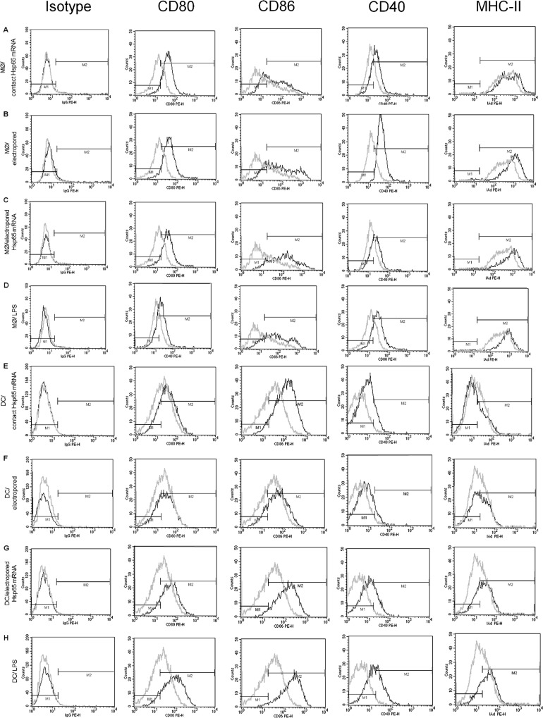
Figure 1. The phenotype of macrophages (A-D) and dendritic cells (E-H) transfected in vitro with Hsp65 mRNA. Macrophages and dendritic cells (DCs) were transfected by contact with Hsp65 mRNA (A and E), electroporated (B and F), electroporated with 5 µg Hsp65 mRNA (C and G) or stimulated with lipopolysaccharide (LPS; D and H). The phenotype of the cells was analyzed by flow cytometry, and the histograms show the expression of the CD80, CD86, CD40, and MHC-II molecules. The gray lines show cells that were not stimulated, and the black lines show stimulated cells. Data are representative of 3 experiments yielding similar results. Mφ = macrophages.

Figure 1.