. 2012 May 25;45(7):583–590. doi: 10.1590/S0100-879X2012007500086

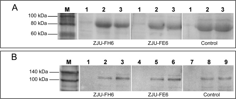
Figure 3. Western blotting and immunoprecipitation analysis of mAbs ZJU-FH6 and ZJU-FE6. A, Western blotting was carried out for cell lysates (50 µg/sample) of the MRC-5, Towne, and AD169 strains. ZJU-FH6 and ZJU-FE6 were used as primary antibody (1 mg/mL, 1:5000). A mouse mAb against gB (Novus Biologicals) was used as a control. Lane 1 = MRC-5; lane 2 = Towne strain; lane 3 = AD169 strain. B, Immunoprecipitation was carried out to immunoprecipitate HCMV gB using ZJU-FH6 and ZJU-FE6 (2 µg/sample). Samples of cell lysates of the Towne and AD169 strains (150 µg/sample) were incubated with ZJU-FH6 (lanes 1, 2, and 3) and ZJU-FE6 (lanes 4, 5 and 6) coupled to protein G-Sepharose beads. A mouse mAb (lanes 7, 8 and 9) against gB (Novus Biologicals) was used as a control. Rabbit anti-human cytomegalovirus gB polyclonal antibodies was used as primary antibody (1:2500). Lanes 1, 4, and 7 = MRC-5; lanes 2, 5, and 8 = Towne strain; lanes 3, 6, and 9 = AD169 strain.

Figure 3.