. 2012 Mar 23;45(5):401–407. doi: 10.1590/S0100-879X2012007500039

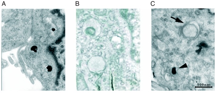
Figure 3. Ultrastructure of PC12 cells examined by transmission electron microscopy 24 h after incubation with phosphate-buffered saline (A), 1-methyl-4-phenylpyridinium (MPP+) (B), and MPP+ plus 3-n-butylphthalide (NBP) (C). In the control and only MPP+-treated groups, PC12 cells barely showed any vacuoles (A, B). In contrast, abundant vacuoles were present in NBP-treated PC12 cells (C). These vacuoles resembled autophagosomes based on the presence of the double-membrane structure (C, arrow). Moreover, single-membrane vesicles, namely autolysosomes, which contained high-density contents, were also observed after NBP treatment (C, arrowhead).

Figure 3.