. 2012 Apr 5;45(6):473–481. doi: 10.1590/S0100-879X2012007500050

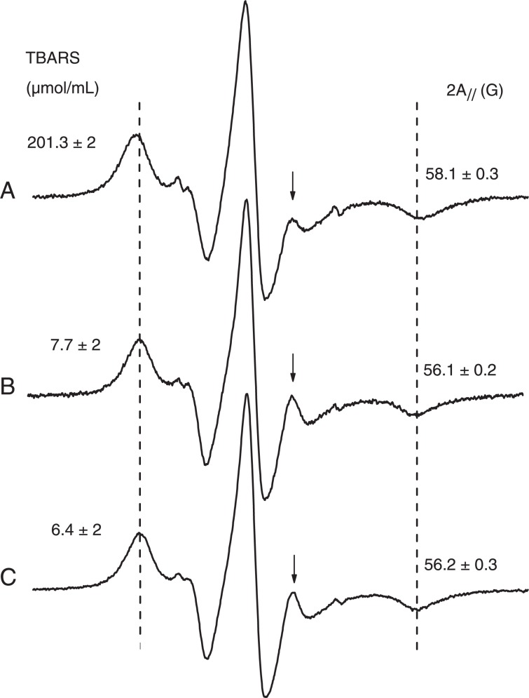
Figure 7. A, Electron magnetic resonance (EPR) spectrum of 5-doxyl stearic acid in erythrocyte ghosts oxidized with 2 mM H2O2 and 200 µM FeSO4. B, Control sample (not oxidized). C, Samples pre-treated with 100 µM (+)-catechin and then oxidized as in A. The EPR parameter, 2A//, increases for the oxidized sample, indicating a reduction in molecular dynamics or increased molecular order. The high-field resonance line of the inner hyperfine splitting (indicated by an arrow for each spectrum) decreases in the spectrum of oxidized membranes, indicating a loss of probe mobility. The figure also shows a decrease in the production of thiobarbituric acid reactive species (TBARS) resulting from lipid peroxidation of erythrocyte ghosts due to the antioxidant activity of (+)-catechin.

Figure 7.