. 2012 Apr 27;45(6):510–516. doi: 10.1590/S0100-879X2012007500061

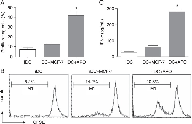
Figure 3. Immature dendritic cells (iDCs) co-cultured with doxorubicin (ADM)-treated apoptotic MCF-7 cells enhance autologous T-lymphocyte proliferation and interferon-γ (IFN-γ) release. Autologous T-lymphocytes were obtained from peripheral blood mononuclear cells of the DC donor by negative selection and were labeled with CFSE dye. Subsequently, the T-lymphocytes were co-cultured with the DCs induced by apoptotic MCF-7 cells (iDC+APO) or untreated MCF-7 cells (iDC+MCF-7) for 5 days. A, The proliferation of T-lymphocytes was assessed by analyzing CFSE dilution by flow cytometry. Data are reported as means ± SD for three independent experiments. B, Representative data from three independent experiments. The percentage of proliferated T-lymphocytes is indicated. C, ELISA was carried out to detect IFN-γ secretion in the supernatants. CFSE = carboxyfluorescein diacetate succinimidyl ester. Data are reported as means ± SD for three independent experiments. *P < 0.05 vs the iDC+MCF-7 group (one-way ANOVA).

Figure 3.