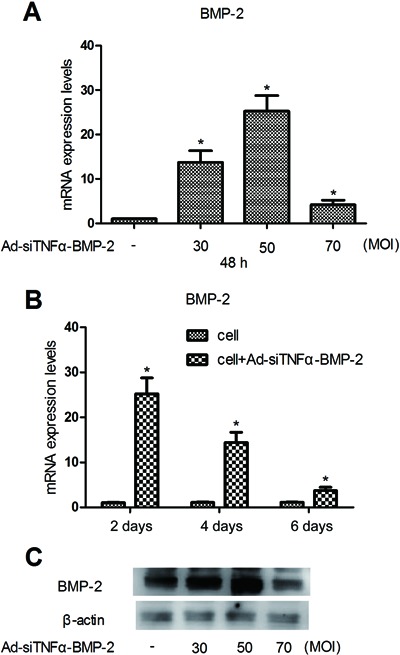
Figure 3. BMP-2 expression in MC3T3-E1 treated with Ad-siTNFα-BMP-2. A, MC3T3-E1 cells were plated on 6-well cluster plates at a density of 4×10⁵ cells/well. After 24 h, MC3T3-E1 cells were treated with Ad-siTNFα-BMP-2 for 48 h. There was a significant increase of BMP-2 mRNA expression. B, Expression of BMP-2 was analyzed at 2, 4, and 6 days in MC3T3-E1 cells treated with 50 MOI Ad-siTNFα-BMP-2. C, BMP-2 protein levels detected by Western blot after 48 h. Data are reported as means±SD. Similar results were obtained in three independent experiments. MOI: multiplicity of infection. *P<0.01, compared to control (one-way ANOVA).

