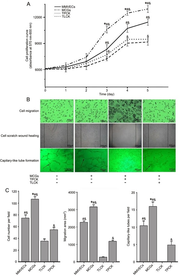
Figure 2. Angiogenesis changes in different conditions in vitro. A, Cell proliferation curve was obtained by AlamarBlue¯ cell viability reagent. B,C, Representative images of migration across Transwell membrane, scratch wound healing and capillary-like tube formation were captured and counted under a contrast phase microscope. Data are reported as means±SE (n=3). MMVECs: myocardial microvascular endothelial cells; MCGs: mast cell granules; TLCK: N-tosyl-L-lysine chloromethyl ketone; TPCK: N-tosyl-L-phenylalanyl chloromethyl ketone. *P<0.05 vs MMVECs, &P<0.05 vs TLCK, #P<0.05 vs TPCK (Student-Newman-Keuls test).