. 2013 Mar 15;46(3):270–277. doi: 10.1590/1414-431X20122551

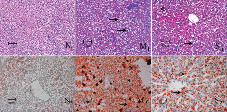
Figure 1. Pathological changes of rat liver stained with hematoxylin-eosin and Sudan III in each group. The micrographs show a mass of large fat granules (black arrows in Panel M1), which represent steatosis in the liver of the group receiving a high-fat diet (HFD) for 6 weeks. Improvement could be seen in the silibinin group with fewer and smaller fat granules (black arrows in Panel S1) and relatively regularly arranged cell cords. Pathologic evaluation showed more and larger fat granules stained salmon pink by Sudan III in the HFD group (black arrows in Panel M2). Fewer and smaller fat granules could be seen in the group treated with silibinin (black arrows in Panel S2). N = control group.

Figure 1