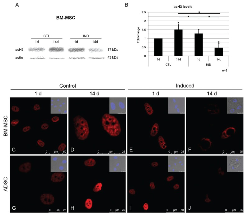
Figure 1. Analysis of histone acetylation during the differentiation of MSCs into adipocytes. A, Western blot showing changes in acH3 levels after 1 and 14 days of control culture conditions (CTL) or adipocyte differentiation induction (IND); an actin probe was used as a loading control. B, Quantitative analyses of relative acH3 levels. Data are reported as means ± SD. MSCs = mesenchymal stem cells; BM-MSCs = bone marrow MSCs; ADSCs = adipose tissue stem cells; d = day. Asterisks just above SD bars show the statistical significance of the difference with respect to control cultures after 1 day. *P < 0.05 (Student t-test). C-J, Immunofluorescence images of BM-MSCs (C-F) and ADSCs (G-J), showing acH3 staining (red) after 1 and 14 days of induction or non-induction. Minor boxes show merged images of differential interference contrast and nuclei counterstained with DAPI.

