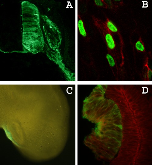
Figure 3. Electroporation results. A, Cross-section view of an electroporated neural tube after cryosections and immunofluorescence detection of enhanced green fluorescent protein (eGFP). The electroporated GFP-positive cells are restricted to the right half of the neural tube and the right dorsal root ganglion (DRG). GFP can also be detected in the commissural neurites that cross to the contralateral neural tube and in the motor neuron projections. Dorsal is up. B, Neural crest cells electroporated with a plasmid encoding the fusion protein Tomato-2A-H2GFP, which is cleaved intracellularly to label the cell membrane with red fluorescence and the cell nucleus with green fluorescence. C, Fluorescence microscope view of a live embryo (embryonic day 3) with the electroporated right lens placode expressing GFP. D, Visualization of GFP (green) and actin (red) in the lens placode after processing cryosections for immunofluorescence with anti-GFP antibody (Abcam, UK) followed by exposure to rhodamine-labeled phalloidin.

