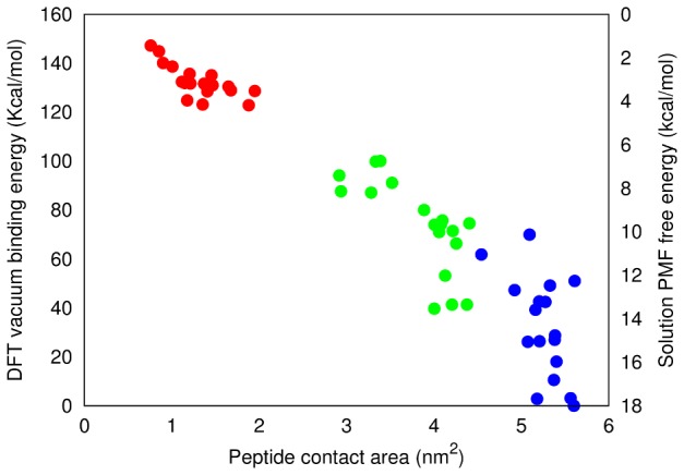
Figure 6. In vacuo binding energies.

Relative DFT vacuum binding energies of apoC-II(60-70) adsorbed to C60 (red), carbon nanotube (green) and graphene (blue) vs the total contact area between the peptide and nanomaterial surface. The solution PMF free energy range for each nanomaterial is also shown (right axis, higher energy = stronger binding) to illustrate the correlation in energies between the classical and electronic structure methods.
