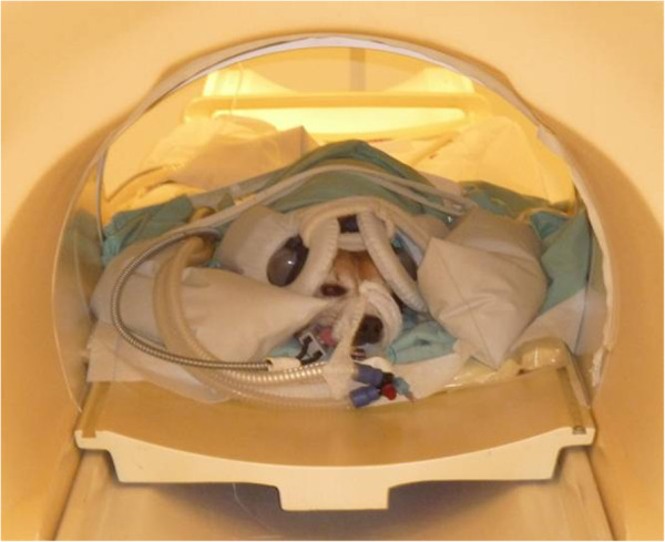
Figure 5.

Examination in the MRI scanner. The dogs were placed in sternal recumbency in the MRI scanner and 11 cm diameter circular surface coils were placed laterally on each side of the dog’s head. Special canine ear covers were used to protect the dog’s hearing and reduce the effects of the background noise.
