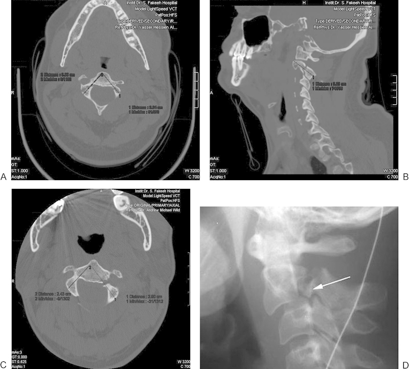
Fig. 2.

(A) Preoperative sagittal computed tomography scan showing the fracture line and the proposed line of screw insertion with the predicted length. (B, C) Axial cuts showing the proposed screw length with inequality of both screws due to anatomical-pathologic variations in both sides. (D) Preoperative plain radiograph of the cervical spine showing C2–C3 subluxation due to fracture at the C2 pars (thin arrow), and the C2–C3 dislocation (thick arrow).
