
Fig. 4.
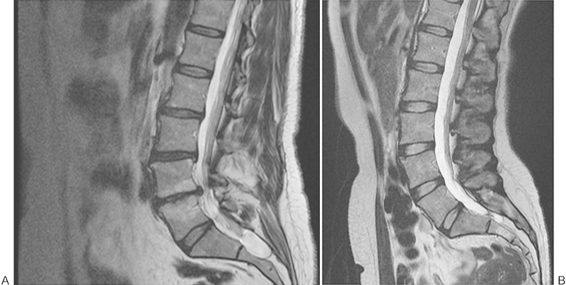
Sagittal T2-weighted magnetic resonance imaging of the lumbar spine. (A) Obese subject with disk degeneration and bulging of L2-L5, with disk space narrowing, end plate irregularities, and Modic changes at L4-L5. Also note the presence of a sacral cyst. (B) A normal-weight individual with nondegenerated lumbar disks.