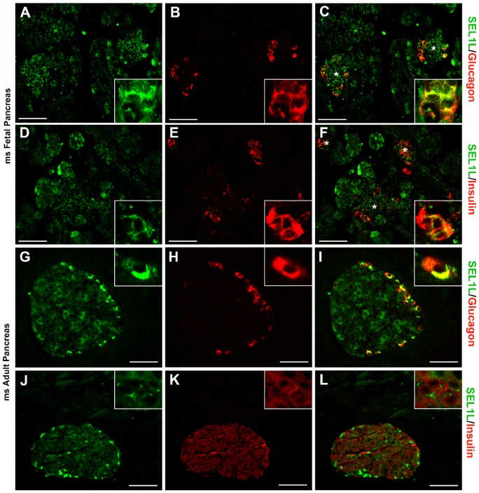
Figure 1. SEL1L expression in fetal and adult mouse pancreas.
Representative images of pancreatic sections from E16.5 mouse embryos (A–F) and 8-weeks-old mice (G–L) immunostained for SEL1L (green; A, D, G and J), glucagon (red; B and H) and insulin (red; E and K). Dual-color immunoflurescence showed SEL1L specific immunoreactivity (green, C and F) in the nascent acinar tissue and in the developing islets (asterisks) stained for glucagon (red, C) and insulin (red, F). While exocrine tissue, in the adult mouse, didn’t show any SEL1L immunoreactivity (green, I and J), endocrine cells revealed a marked expression of SEL1L protein with a strong cytoplasmic immunoreactivity in α-cells (stained for glucagon in red, I) and a moderate expression in β-cells (stained for insulin in red, L). Scale bar = 50 µm.

