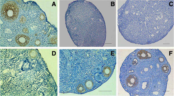
Figure 6.

AMH expression in recipient mouse ovaries. (A) AMH was expressed in all granulosa cells of primary, preantral and small antral follicles in normal ovaries. (B) AMH expression disappeared in stromal and atretic follicles in recipients ovaries without hAECs transplantation two months after chemotherapy. (C) AMH expression was negative in recipient ovaries seven days after hAEC transplantation. However, AMH expressions were detected in recipient ovaries after hAEC transplantation for 14 days (D) and 21 days (E). (F) AMH expression patterns are strong in recipient ovaries two months after hAEC transplantation; data are consistent with that from control normal ovaries. Scale bars = 100 μm. AMH, Anti-Müllerian hormone; hAEC, human amniotic epithelial cell.
