Figure 2.
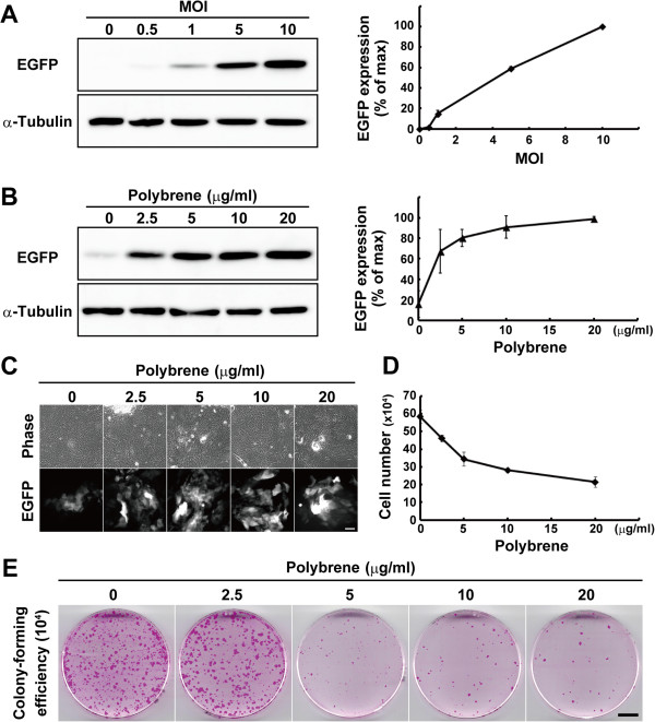
The effects of polybrene on gene transduction efficiency and growth potential of keratinocytes. (A) The left panel shows Western blotting of lysates from keratinocytes transduced with the enhanced green fluorescent protein (EGFP)-expressing lentivirus at various multiplicity of infection (MOI). The expression of α-tubulin was used as loading control. Right panel shows relative expression levels of EGFP. The values (mean ± SD) were obtained from the density of bands normalized with α-tubulin bands. (B) The left panel shows Western blotting of lysates from keratinocytes transduced with the EGFP-expressing lentivirus at MOI 1, and various concentration of polybrene. The expression of α-tubulin was used as loading control. Right panel shows relative expression levels of EGFP. The values (mean ± SD) were obtained from the density of bands normalized with α-tubulin bands. (C) Images of phase-contrast and EGFP expression in keratinocytes transduced with the EGFP-expressing lentivirus at MOI 1, and various concentrations of polybrene. Bar, 100 μm. (D) The effects of polybrene on keratinocyte proliferation are shown. Keratinocytes (104 cells) were cultivated with various concentration of polybrene during lentivirus transduction, and the number of keratinocytes were counted after seven days of infection. (E) Determination of colony-forming efficiency (CFE) of keratinocytes infected with lentiviral particles at MOI 1, and various concentrations of polybrene. After seven days of infection, 104 cells were passaged and cultured without polybrene for eight days until the cultures were fixed and stained with rhodamine B. Bar, 10 mm. The values (means ± SD) in A, B, and D were determined based on results from triplicate experiments.

