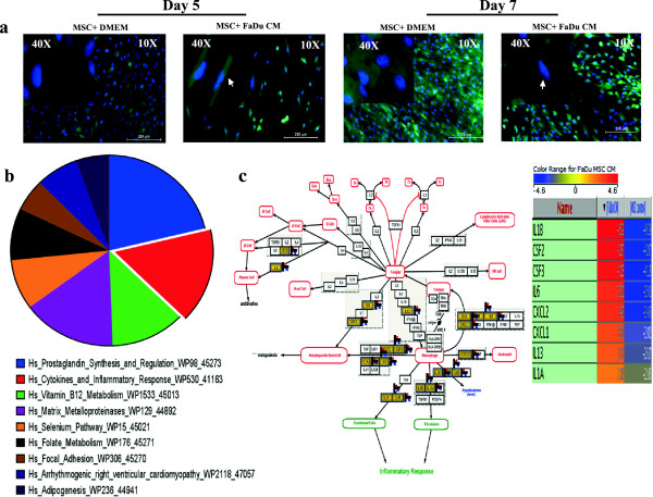
Figure 1.
Effects of FaDu conditioned medium (CM) on human MSC morphology and gene expression. (a) Representative micrographs of MSC-GFP cells grown under normal conditions (left panel) or exposed to FaDu CM (right panel). Hoechst 33342 was used for nuclear staining and images were obtained at the indicated time points (10× magnification, 200 μm scale bar). Arrow heads point to MSCs with fibroblastic morphology in CM treated cells. (b) MSCs grown under normal conditions or exposed to FaDu CM were subjected to microarray analysis. Differentially upregulated genes in MSCs exposed to FaDu CM were subsequently subjected to pathway analysis as described in Methods. The pie chart represents the top ten pathways where the pie size represents percent enrichment of the pathway. (c) Genes in the cytokine and inflammatory response pathway were among the most highly enriched category in the microarray data. MSCs, mesenchymal stem cells.

