
Figure 2.
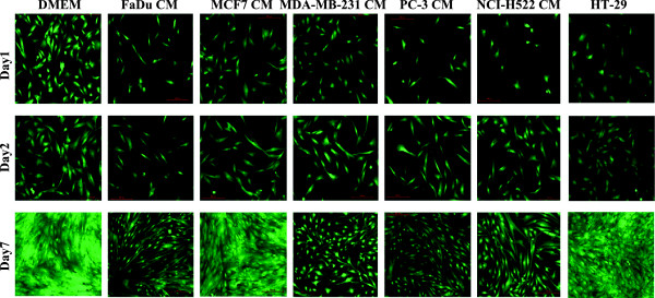
Comparative analysis of morphological changes in MSCs exposed to conditioned medium (CM) from a panel of human cancer cell lines. MSCs were grown under normal conditions ((D)MEM) or were exposed to CM from the indicated cancer cell lines (FaDu, MCF7, MDA-MB-231, PC-3, NCI-H522 and HT-29) and, subsequently, images were obtained on days1, 2 and 7. Representative micrographs from at least three independent experiments are shown. All images were taken using 10x magnification. MSCs, mesenchymal stem cells.