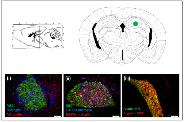
Figure 1.

Characterisation of responses following mesenchymal stem cell transplantation. The upper panel of Figure 1 demonstrates a transveral and coronal brain slice at the site where mesenchymal stem cells (MSCs) were transplanted (green dot). The bottom row of Figure 1 demonstrates immunofluorescent pictures of cryosections from FVB mice transplanted with 2 × 10⁵ enhanced green fluorescent protein (eGFP)- and luciferase-expressing MSCs into the striatum. (i) Transplanted MSCs are recognised by the brain’s immune system, demonstrating Iba1+ microglia (blue) invading and activated GFAP + astrocytes (red) surrounding the eGFP-expressing MSC implant (green) at week 1 post-implantation. (ii) A proportion of the Iba1+ microglia are classically activated and demonstrate CD11b (blue) and NOS2 (red) expression, corresponding to a neuroinflammatory microglial phenotype. (iii) At 6 h post-transplantation a lot of hypoxic (red) cells are found within the eGFP-positive (green) MSC transplant, leading to death of about half of the transplanted cells. The representative pictures were taken at a magnification of 20 × .
