Figure 2.
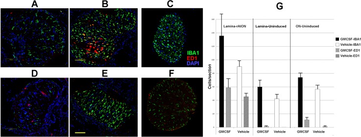
Intraventricular GM-CSF increases inflammation in the infarcted ON. Confocal photos show inflammatory cells in representative sections of lamina (first 500 μm) and more distal (>2 mm) ON regions of the different treatment groups. (A–C) GM-CSF-treated animals. (D–F) Vehicle treated animals. (A, D) Uninduced lamina sections. Intrinsic microglial cells (IBA1[+], in green) are nonactivated with an extended/protoplasmic appearance). The majority of ED1(+) cells (in red) are present in vessels, with few ED1(+) cells present in the ON. (B, E) rAION-induced lamina sections. There is extensive microglial activation, and ED1(+) systemic macrophage invasion is seen in sections. (C, F) Uninduced distal ON sections. Few ED1(+) cells are present. The uninduced GM-CSF-treated ON (C) has microglial activity similar to that seen in the vehicle-treated (F) uninduced nerve. (G) Quantification of lamina/ON tissue sections from three individuals. The rAION-induced, GM-CSF-treated lamina shows a trend towards the greatest number of microglia and systemic macrophages, compared to the laminae from rAION-induced, vehicle-treated animals. This trend is continued in GM-CSF-treated tissues from uninduced eyes. Scale bars: 100 μm (B, E).

