Figure 5.
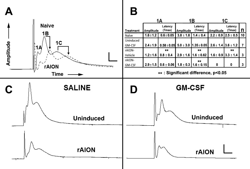
Rodent NAION results in postinfarct demyelination/myelin dysfunction. Ex vivo electrophysiological analysis using CAPs. (A) Comparison between contralateral naïve and rAION induced ONs (same animal comparisons). The ON-CAPs reveal three myelinated axonal components: Large (1A), medium (1B), and small (1C) diameter fibers. At 1 month post-rAION in untreated (rAION only, no intraventricular treatment), there is a considerable decrease in the amplitudes of all three components, while 1B and 1C fibers also show reduced transmission speed, consistent with postinfarct demyelination. (B) Quantification of CAP parameters in representative animals. The last column on the right indicates the number of animals tested per group. Vehicle-treated animals show a reduction in amplitude and delayed transmission speed in all three fiber sizes in the induced eyes 1 month after rAION. The GM-CSF-treated animals also reveal decreased amplitude and reduced transmission speed of the largest fibers, loss of amplitude in the mid-size fibers, and a complete dropout of the smallest (1C) myelinated fiber component. (C, D) Qualitative comparison of ONs from (C) vehicle- and (D) GM-CSF-treated animals. There is decreased 1A and 1B fiber amplitude, and 1C fiber transmission speed delay in the vehicle-treated animal. There is total loss of the 1C fiber component in the rAION-induced, GM-CSF-treated ON. The 1A peak is increased in amplitude, while the 1B peak is reduced in amplitude. Scale bars: 1.5 mV and 0.75 msec (A), 3 mV and 1.5 msec (C, D).

