Figure 6.
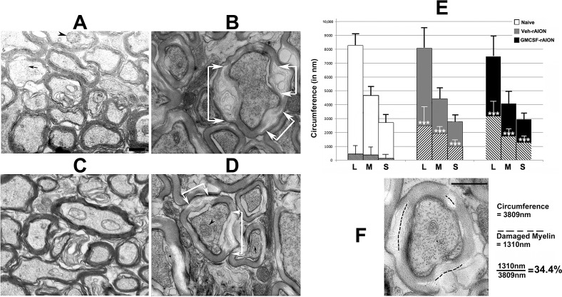
Rodent NAION results in long-term focal myelin damage. (A) Uninduced (naïve) ON. Intact axons of varying calibers are packed together, with normal myelin. Few disruptions are discernible. (B) Vehicle-treated, rAION-induced. While axoplasm is intact with mitochondria and neurofilaments, focal regions of myelin damage and swelling are apparent (areas indicated by white arrows). (C) GM-CSF-treated, uninduced. Axonal structure is similar to that seen in (A). (D) GM-CSF-treated, rAION-induced. Similar to (B), there are focal areas of myelin degeneration and swelling, indicated by white arrows. (E) Myelin damage quantification in naïve, vehicle-rAION and GM-CSF-rAION–induced animals (n = 10 axons of each caliber for each group). The method of determining axonal size by circumference and myelin damage for each axon is shown in (F). In (D), three axonal fiber sizes (L, large; M, medium; S, small) are revealed by circumferential measurement. Hatched areas within each larger bar represent mean myelin damage. Few naïve (white bars) axons of any size show myelin damage. By contrast, vehicle-treated rAION-induced (gray bars) axons and GM-CSF-treated rAION-induced (black bars) axons of all sizes show significant levels of myelin damage (***P < 0.01; 2-tailed t-test) compared to that seen in their naïve equivalent fiber size. The GM-CSF-treated ONs show a nonstatistically significant trend towards more myelin damage. Scale bars: 500 nm (A, F).

