
Figure 2. A novel DNA insertion mutation within the Ppd genetic interval.
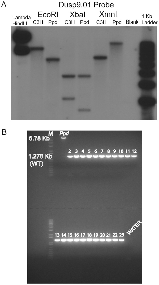
A. Southern blot demonstrating abnormal product sizes in male Ppd genomic DNA compared to male C3H DNA hybridized with Dusp9.01 probe. Southern blot comparisons with other strains revealed these alterations to be Ppd-specific (Figure S1). B. The Ppd insertion is not present in 21 different mouse strains and CD-1 (original strain). X-chromosome interval-specific PCR primers (F1/R2, see Figure 3) were used to amplify affected male Ppd DNA compared with male genomic DNA samples (obtained independently from Jackson Labs) from the following strains: Lane 2: 129S1/SvImJ; 3: 129X1/SvJ; 4: A/J; 5: AKR/J; 6: Balb/cByJ; 7: Balb/cJ; 8: C3H/HeJ; 9: C57BL/6J; 10: C57BL/10J; 11: CAST/EiJ; 12: CBA/J; 13: CD10/JlsJ; 14: CZECHII/EiJ; 15: DBA/1J; 16: DBA/2J; 17: FVB/NJ; 18: MOLF/EiJ; 19: MSM/Ms; 20: SJL/J; 21: SPRET/EiJ; 22: SWR/J. Representative outbred CD-1 (Charles River Labs) genomic DNA is shown in lane 23 adjacent to the water control. Analysis of 11 other independent male CD-1 mouse DNA samples also revealed only the 1.28 kb wild-type PCR product (not shown). The 6.8 kb product spanning the insertion was only observed using Ppd DNA; the 1.28 kb wild-type product is labeled.