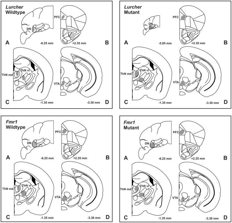
Fig. 1.
Representative coronal sections in Lurcher and Fmr1 wildtype and mutant mice illustrating placements (gray shaded areas) of (A) stimulating electrodes in the dentate nucleus (DN), (B) dopamine recording electrodes in the medial prefrontal cortex (mPFC), (C) infusion cannulae in mediodorsal thalamus (ThN md) and ventrolateral thalamus (ThN vl), and (D) infusion cannulae in the ventral tegmental area (VTA). Numbers correspond to mm from bregma. Placements of stimulating and recording electrodes, and cannula placements overlapped in all groups. Sections were adapted from the mouse atlas of [37].

