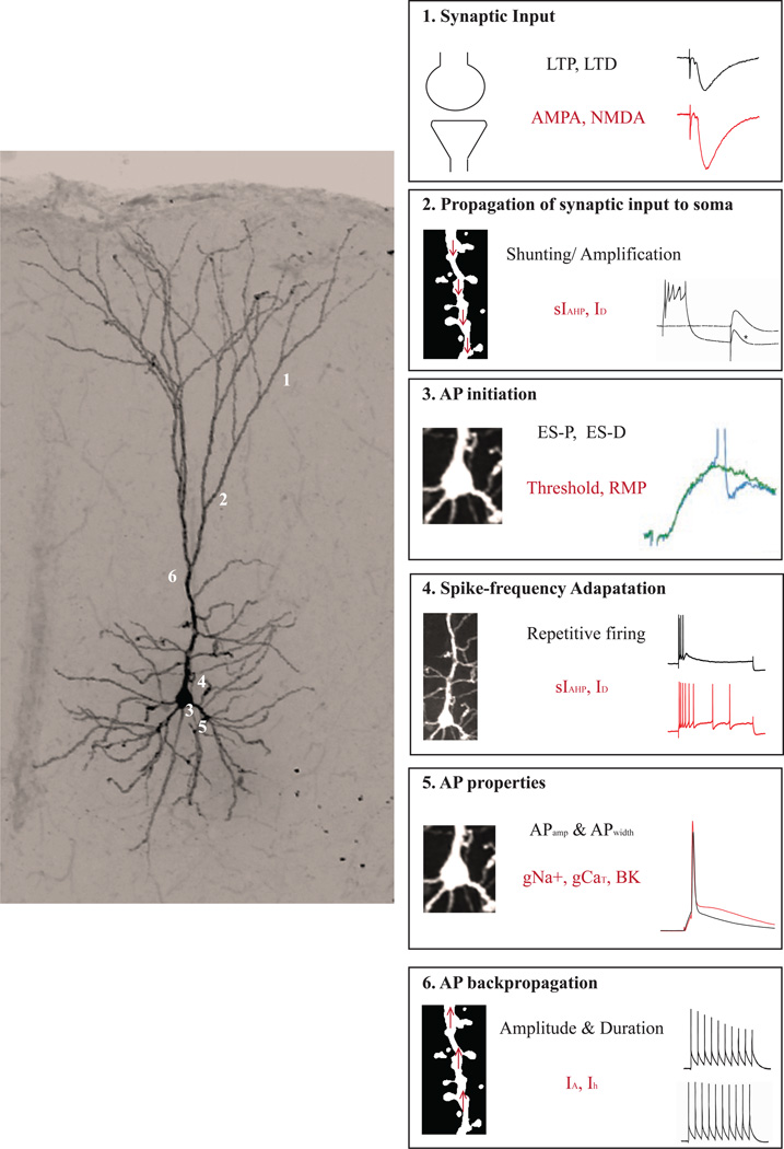
Figure 1. Synaptic and intrinsic properties shape neuronal information processing.
Left panel depicts a medial prefrontal cortical neuron filled with biocytin during whole-cell patch clamp recording and imaged using confocal microscopy (Olympus FV1200). Numbers 1, 2, 3, 4, 5 and 6 refer to the boxes in the right panel. (1) A vast majority of neuronal input originates on the dendritic spines with smaller contributions from synapses that are made on the dendrites, soma and axons. Synaptic inputs can undergo bidirectional plasticity in the form of LTP and LTD by modulation of AMPA and NMDA receptor-mediated transmission. (2) Propagation of the synaptically generated signal (EPSP) depends upon the active and passive dendritic properties including ionic conductances that contribute to the afterhyperpolarization (AHP). (3) Once the signal reaches the soma, neuronal output is determined based on factors like AP initiation threshold, resting membrane potential, etc…, which in turn rely on ion channels within the soma. (4) Bidirectional plasticity impacting the coupling of EPSPs to spikes is referred to as ES-P and ES-D. The number of APs generated following sustained stimulation (spike frequency adaptation) can also code relevant information, and relies upon K+ conductances, including those that underlie the AHP. (5) In addition, properties like amplitude and duration of APs can also modulate pre- and postsynaptic aspects of neuronal processing like neurotransmitter release and bAPs. (6) The magnitude and travel distance of bAPs can be influenced by IA currents, which can ultimately modulate Ca2+ influx into the dendritic compartment. Abbreviations: long term potentiation, LTP; long term depression, LTD; excitatory postsynaptic potential, EPSP; afterhyperpolarization, AHP; action potential, AP; EPSP-to-Spike coupling potentiation, ES-P; EPSP-to-Spike coupling potentiation, ES-D; backpropagating APs, bAPs. Electrophysiological traces in boxes 2, 3, 5 and 6 were adapted from Sah and Bekkers (1996), Daoudal et al. (2002), Deng et al. (2013) and Tsubokawa et al. (2000) respectively, with permission.

