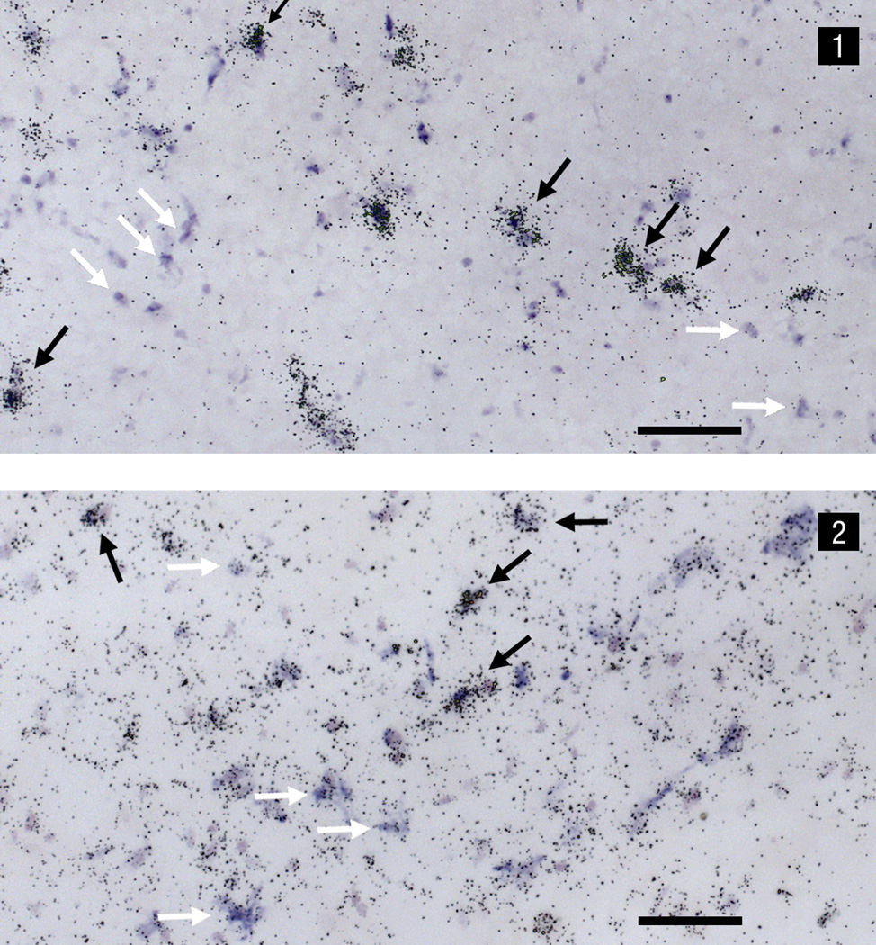
Fig.C.
Dual in situ hybridization photomicrographs under bright-field illumination of (1) FGF-R1-35S mRNA and CCK-digoxigenin mRNA, and (2) CCK-35S mRNA and FGF-R1-digoxigenin mRNA. Labeling indicating the presence of the non-radioactive digoxigenin probe appears as a blue-purple precipitate, whereas the smaller, punctate silver grains are indicative of the radioactive 35S probe. Black arrows indicate cells that express both CCK and FGF-R1 mRNA. White arrows indicate separate digoxigenin-labeled CCK mRNA (1) and 35S-labeled CCK mRNA (2) cells. Scale bar = 100µm.

