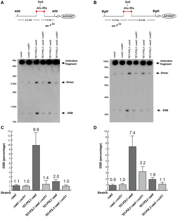
Figure 4. Detection of DSB accumulation in wild-type and mutant strains upon deletion of RAD51.
DSB detection was carried out as described in Figure 2. The strains used in this analysis are: Δsae2, Δsae2Δrad51, TET-POL3Δsae2, TET-POL3Δsae2Δrad51, TET-POL2Δsae2, TET-POL2Δsae2Δrad51. (C) and (D) Densitometry analysis of the broken fragments normalized to the intact chromosome V in Δsae2 strains in (A) and (B), respectively. Values are shown as mean (shown on the top of the bars) with standard deviation obtained from at least three independent experiments.

