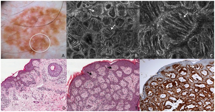
Figure 1. Superficial spreading melanoma in situ.
This lesion shows on dermoscopy (A) a slightly pigmented network (white circle corresponds to the punch area). RCM mosaic image (B, 1×1 mm) at the level of the DEJ shows irregular and dishomogeneous dermal papillae with dendritic cells (white arrows). RCM individual image (C, 0,5×0,5 mm) at the level of the DEJ shows dendritic cells forming “bridges” called mitochondria-like structures (white arrow). Perpendicular section (D) shows disarrangement of the rete ridge and the increased number of atypical melanocytes. Transverse section (E, HE staining) shows atypical melanocytes protruding into the dermal papillae forming bridges (black arrows). Transverse section (F, Melan-A staining) shows cells positive for Melan-A protruding into the dermal papillae (white arrows).

