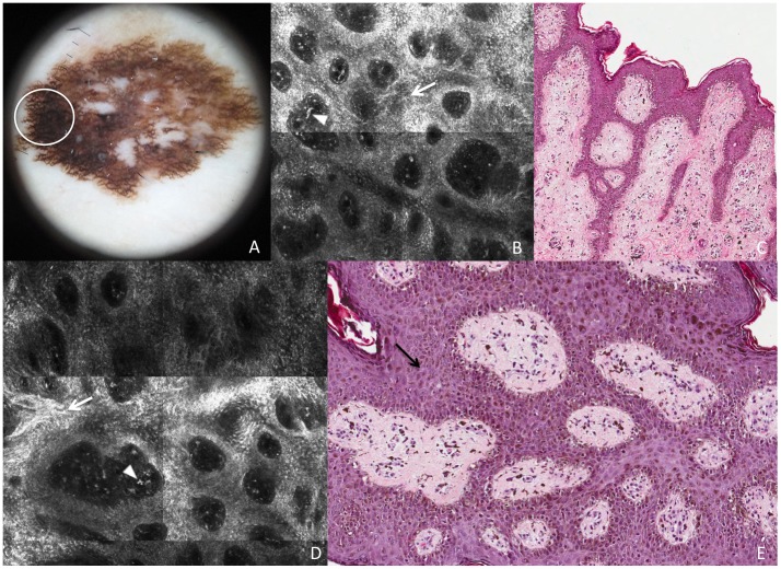
Figure 2. Superficial spreading melanoma in situ.
This lesion shows on dermoscopy (A) a broadened pigmented network (white circle corresponds to the punch area). RCM mosaic images (B and D, 1×1 mm) at the level of the DEJ show demarcated and non-demarcated rings separated by loosely thick interpappilary spaces (white arrows) and some plump bright cells and bright dots are visible within dermal papillae (arrowheads). Perpendicular section (C) shows disarrangement of the rete ridge and the increased number of atypical melanocytes in the epidermis. Transverse section (E) shows predominance of atypical melanocytes, isolated or in nests, enlarging the interpapillary spaces (black arrow).

