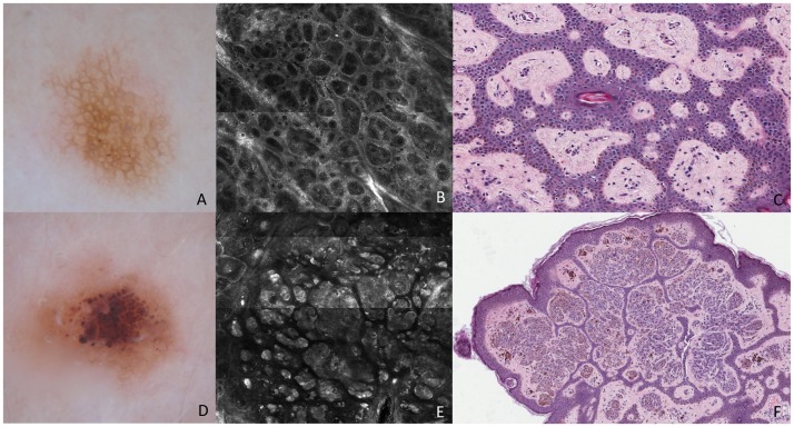
Figure 3. Melanocytic Nevi.
These lesions show typical network (A) and regular globules (D), on dermoscopy. RCM mosaic image (B, 1×1 mm) at the level of the DEJ shows rings of bright polygonal cells surrounding roundish to oval dark areas corresponding to dermal papillae at DEJ. Transverse section (C) shows isolated melanocytes arranged around the dermal papillae and there are nevus cells nests within the epidermis. RCM mosaic image (E, 1,5×1,5 mm) at the level of the DEJ and dermis shows compact aggregates of large polygonal cells similar in morphologic features and reflectivity, forming polyhedral structures. Transverse section (F) shows dense nests composed of nevus cells within the dermis surrounded by a narrow band of epidermis.

