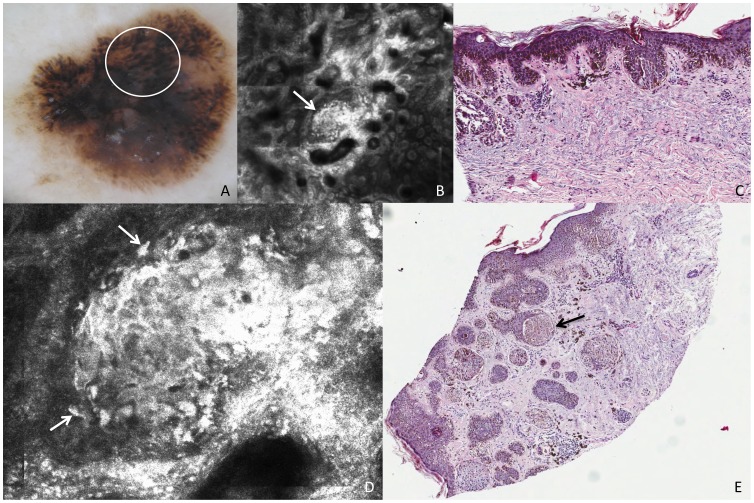
Figure 4. Superficial spreading melanoma, Breslow thickness 0,8 mm.
This lesion shows on dermoscopy (A) irregular globules (white circle). RCM mosaic image (B, 1,5×1,5 mm) at the level of the DEJ shows irregularly shaped clusters (white arrow). Perpendicular section (C) shows compact aggregates of atypical melanocytes with a slight intercellular discohesion. RCM individual image (D, 0,5×0,5 mm) at the level of the DEJ shows a cluster with cells that are nonhomogeneous in morphologic features and reflectivity (white arrows). Transverse section (E) shows large amount of atypical melanocytes predominantly in nests with variable size and shape within the epidermis and the dermis. The black arrow points to the nest that makes the exact correlation with confocal image.

