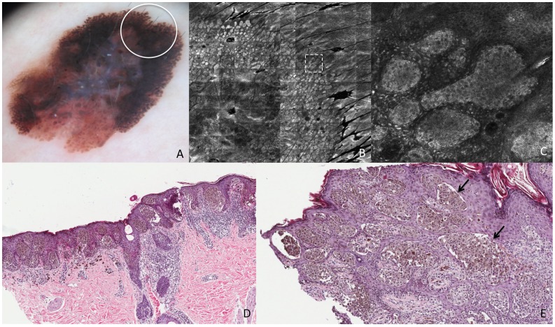
Figure 5. Superficial spreading melanoma, Breslow thickness 0,79 mm.
This lesion shows on dermoscopy (A) pseudopods (white circle corresponds to the punch area). RCM mosaic image (B, 3×3 mm) at the level of the DEJ shows compact aggregates of atypical cells distributed in a linear arrangement toward the periphery with a dense nest at the extremity (the area inside the dashed square is represented in figure C). RCM individual image (C, 0,5×0,5 mm) at the level of the DEJ shows a pseudopod in detail, characterized by elongated, dense and bright peripheral aggregate. Perpendicular section (D) shows nests of atypical melanocytes distributed contiguously toward the periphery along the DEJ. Transverse section (E) shows nests of atypical cells arranged in a linear manner throughout the periphery of the lesion (black arrows).

