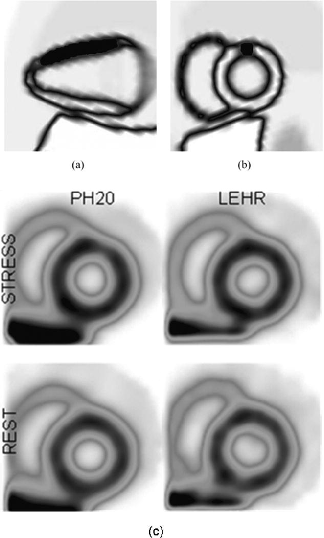
Figure 8.

(a) Perfusion defect geometry (20% contrast). (b) Short axis view of the phantom with perfusion defect in (a). (c) Reconstructed PH20 and LEHR short-axis slices of the phantom in (a). An abnormality is present in the PH20 images. No abnormality is apparent in the LEHR images.
