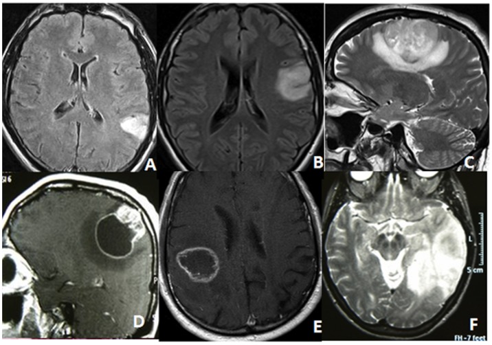
Figure 1. These preoperative MRIs depict the five classes of subcortical infiltration pattern.
(A) class 1: tumors invading and confined to only 1 gyrus without infiltration of white matter connections; (B) class 2: tumors invading 1 gyrus with extension to white matter and/or adjacent gyrus; (C) class 3: tumors infiltrating up to 3 gyri and extending toward the long range white matter tracts; (D) the same as class 3 but with a large cystic component; (E) class 4: tumors primarily located in the white matter under eloquent gyri; (F) class 5: lobar tumors.

