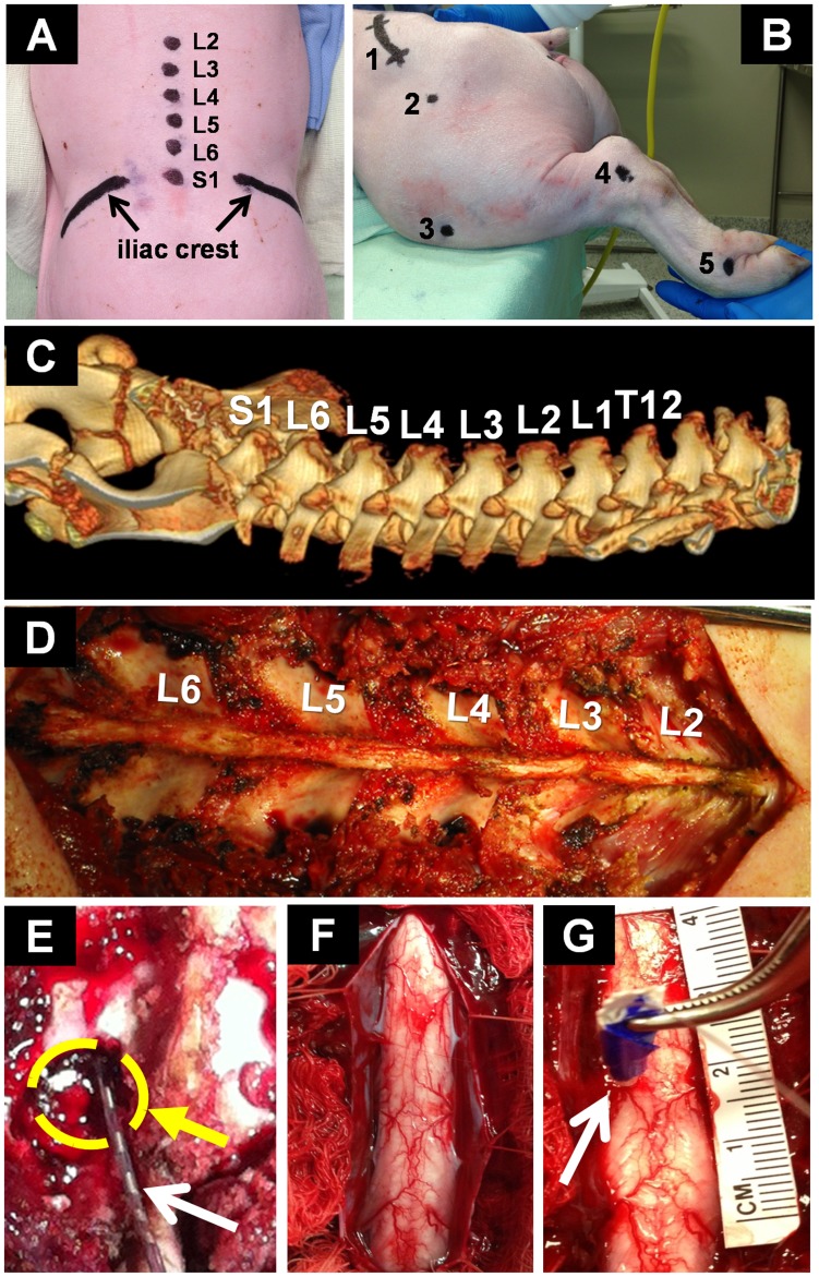
Figure 1. Experimental setup.
(A) Anatomical landmarks (i.e., sacrum, iliac crest, and spinous processes) for localization of the lumbar spine (L2–S1); (B) Lateral view of anatomical motion analysis markers (1-Lateral iliac crest, 2-Trochanter major, 3-Patella, 4-Lateral malleolus, 5-Fourth metatarsal); (C) Pre-operative computed tomography: Lower thoracic vertebrae, lumbar vertebrae and sacrum; (D) Exposure of the lumbar spine; (E) Laminotomy at L5-left (circle) and epidural electrode (arrow); (F) Exposure of the spinal cord for intraspinal microstimulation; (G) Typical microelectrode implantation in the left hemicord (arrow). Insulating adhesive tape was used for protection of the electrode during forceps-insertion and color-coded to determine insertion depth (e.g., blue tape corresponded to an electrode depth of 6 mm). A reference ruler (centimeters) was used to determine cord dimensions and estimate electrode location.

