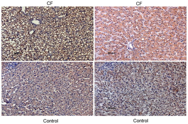
Fig. 3.
Immunohistochemical localization of SULT1E1 in human pediatric CF liver. Paraffin sections of liver from children with CF and age-matched controls were treated with a microwave antigen retrieval technique and incubated with rabbit anti-SULT1E1 IgG. After labeling with the biotin–streptavidin complex and counter-stained with hemotoxylin, visualization was carried out with DAB. Stained sections were photographed at 600× magnification. The upper panels are from CF patients and the bottom panels from controls. The upper-left panel (9 mo F) had steatosis and the highest staining, the upper-right panel was from a 4 yo F. The lower panels were from 9 mo M (left) and 4 yo F (right).

