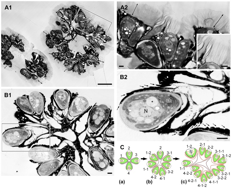
Figure 2. Colony of B. braunii race B by electron microscopy.
A. Sample frozen in liquid propane, B. Sample frozen by high-pressure freezing machine. A1. Each colony is enclosed by colony sheath (double ended arrow). A2. Enlargement of the rectangle in A1. Colony sheath is composed of fibrils stretching from the apical region of each cell and the upper edge of the electron-dense intercellular matrix. Inset is the enlargement of a part of A2. B1. Each cell is covered with 3–6 electron-dense thin layers in the basolateral region. These layers are appeared to be holding the colonial cells together. B2. Enlargement of the rectangle in B1 (small arrow indicates each electron-dense thin layer). C. A speculative genealogic relationship among the cells in the colony shown in B1. ch, chloroplast; N, nucleus; *, lipid body. Scale bars in A1 and A2–B2: 10 µm and 1 µm, respectively.

