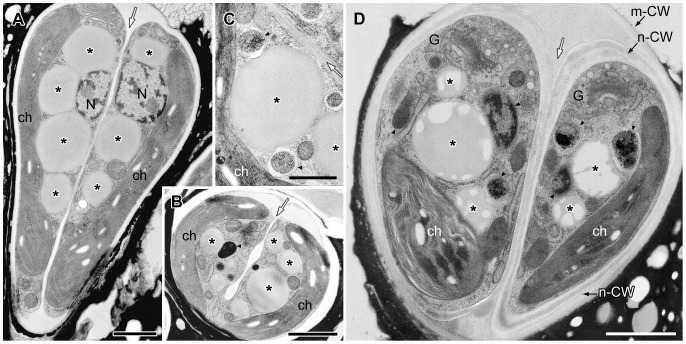
Figure 5. EM analysis of lipid bodies after septum formation to just completing cell wall formation.
A. Longitudinal section of dividing cells just after septum formation but before cell wall formation. B. Cross section of septum developing cell. C. Lipid bodies contact to electron-dense bodies enclosed unit membrane. D. A pair of daughter cells during cell wall formation. Note that all lipid bodies in the earlier stage (A) are filled with a similar electron-dense material but in D they became patchy with emergence of electron-dense bodies. ch, chloroplast; G, Golgi body; m-CW, mother cell wall; N, nucleus; n-CW, new cell wall; arrowhead, electron-dense body enclosed by unit membrane; white arrow, septum; * lipid body; arrowhead, electron-dense body enclosed by unit membrane. Scale bars in A,B,D and C: 1 µm and 0.5 µm, respectively.

