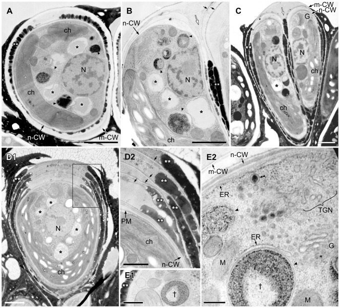
Figure 6. EM analysis of lipid bodies in a pair of daughter cells accumulating oil droplets.
Between a new cell wall and a mother cell wall at the basolateral region, oil droplets arrange side by side and form 1-6 layers; one layer in A, E, two layers in B, C, and five layers in D are observed. A–C. Lipid bodies contact to electron-dense bodies enclosed unit membrane. D. There is a thin layer between oil droplets layers. D2. Enlargement of the rectangle in D1. E. Cell accumulates one oil droplet layer. E2. Enlargement of a part of E1, in which electron-dense body enclosed by unit membrane marked with † is same marked one in E1. The rER lacks ribosomes on the surface facing the plasma membrane or electron dense body. ch, chloroplast; ER, endoplasmic reticulum; G, Golgi body; m-CW, mother cell wall; M, mitochondrion; N, nucleus; n-CW, new cell wall; PM, plasma membrane; TGN, trans-Golgi network; * lipid body; **, oil droplet on the cell surface; small arrow, thin layer between oil droplets layers; white arrow, septum; arrowhead, electron-dense body enclosed by unit membrane; double arrowheads, Golgi vesicle. Scale bars in A-D1, D2-E1 and E2: 1 µm, 0.5 µm, and 0.2 µm, respectively.

