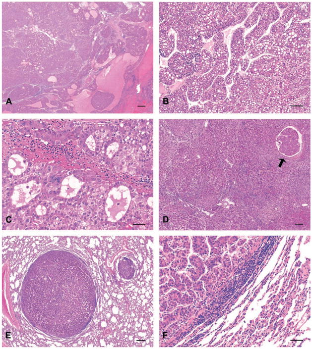
Figure 1.
Hepatocellular carcinomas (HCC), rhesus macaques exposed to diethylnitrosamine (DENA), hematoxylin and eosin (H&E) stained tissue sections. (A) primary HCC, macaque 1102P liver. Hepatic architecture has been effaced by diffuse hepatocellular carcinoma accompanied by hemorrhage and necrotic foci. Bar = 500 μm. (B) Primary HCC exhibiting trabecular arrangement, macaque 1236T liver. Bar = 100 μm. (C) Primary HCC exhibiting pseudoglandular arrangement, macaque 1088P liver. Bar = 50 μm. (D) Primary HCC exhibiting solid pattern of growth with occasional trabecular arrangement, macaque 595G liver. Note focus of cancer invasion within hepatic blood vessel (arrow). Bar = 250 μm. (E) Pulmonary metastatic HCC exhibiting solid growth pattern with occasional trabecular arrangement, macaque 595G lung. Bar = 300 μm. (F) Metastatic HCC exhibiting trabecular arrangement observed in portions of the primary cancer illustrated in the corresponding primary HCC (D), macaque 595G lung. Bar = 50 μm.

