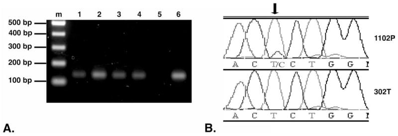
Figure 3.

Detection of β-catenin mutation in hepatocellular carcinoma (HCC). (A) Polymerase chain reaction (PCR) amplification of β-catenin. PCR was carried out using genomic DNA isolated from paraffin-embedded tissues. PCR products were separated by electrophoresis on a 1.5% agarose gel. Lanes: (m) 100 bp DNA ladder, representative 130 bp amplified DNA product from unexposed and diethylnitrosamine (DENA)-exposed macaque livers: (1) 302T, (2) 4T, (3) 595G, (4) 1102P, (5) no DNA template negative control, and (6) genomic DNA isolated from FRhK4, a rhesus macaque kidney cell line (CRL-1688, American Type Culture Collection, Manassas, VA), used as a positive control. (B) Partial sequence chromatograms depicting nucleotide sequences of the β-catenin gene exon 3 in liver tissue of DENA-exposed macaque 1102P (top row), compared to sequence from colony breeder rhesus macaque 302T (bottom row). The T → C missense mutation (arrow) resulted in a Ser → Pro mutation at amino acid 33.
