
Figure 3.
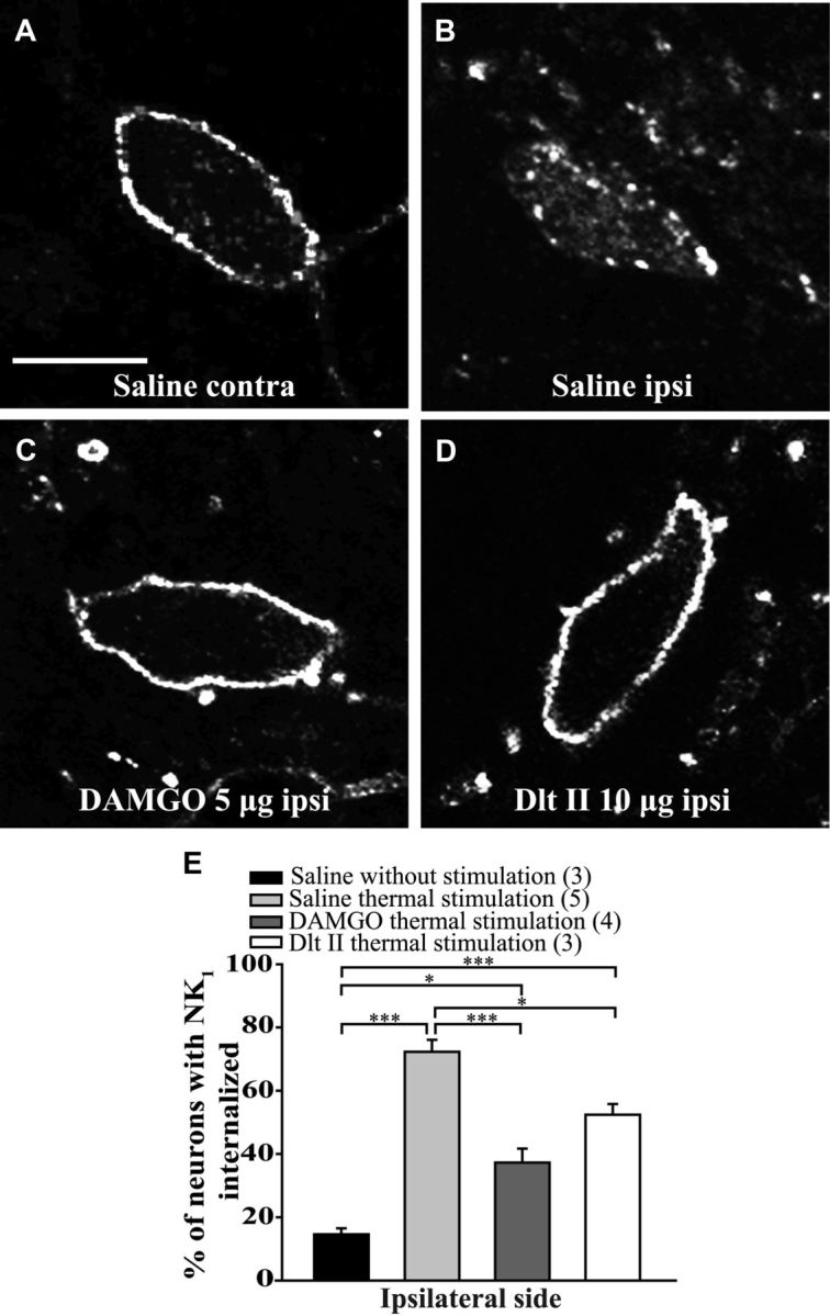
Intrathecal DAMGO and Dlt II reduce heat-induced NK1 receptor internalization. Internalization of NK1 receptors was induced by immersing the right hindpaw of male Sprague Dawley rats in a 49°C water bath for 38 s, and lamina I NK1 receptor-immunoreactive neurons were observed by immunofluorescence. The noxious heat stimulation was applied 10 min after intrathecal injection of saline (A,B), DAMGO 5 μg (C), or Dlt II 10 μg (D). Confocal images of neurons on the contralateral (A) and ipsilateral (B–D) sides of the spinal cord are shown. On the contralateral side of the saline-injected animals, immunolabeling of NK1 receptors appeared to be at the cell surface (A). However, on the ipsilateral side of the same animals, the noxious heat stimulation induced a significant increase in NK1 receptor internalization, as evidenced by the intensely labeled intracellular vesicle-like structures (B). When DAMGO (C) or Dlt II (D) was injected, a significant reduction in NK1 receptor internalization was observed on the ipsilateral side compared with the same side in saline-injected rats. The animals that had received a saline injection but no noxious stimulation had low basal proportions of neurons with internalized NK1 receptors. This result is illustrated in the graphic representation showing the percentage of neurons with NK1 receptor internalization induced by heat stimulation for ipsilateral side of the lumbar spinal cord (E). *p < 0.05 (one-way ANOVA with Bonferroni's post hoc test). ***p < 0.001 (one-way ANOVA with Bonferroni's post hoc test). The numbers in parentheses represent the number of animals per group. Error bars indicate the SEM. Scale bar: A, 30 μm.