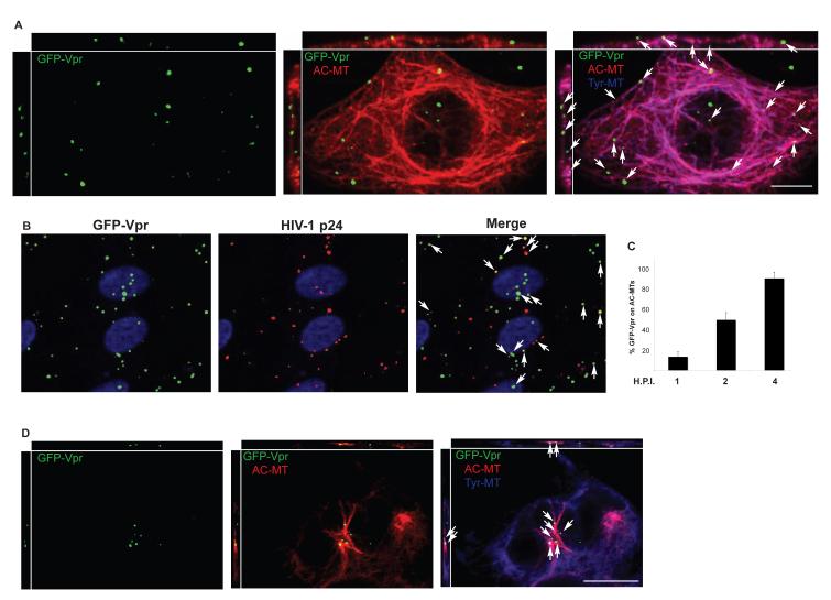
Figure 3. HIV-1 localizes to stable MTs in the presence of nocodazole.
(A) CHME3 cells were infected with HIV-1-VSV-GFP-Vpr at m.o.i. 1 for 4h. Cultures were fixed and stained for Tyr-MTs (blue), AC-MTs (red) and GFP (green). Arrows point to viral particles localized to AC MT networks. Orthogonal views are presented above and to the left of each regular imaging plane (top x-z, left z-y), scale bar, 10μm. (B) Cells were infected with HIV-1-VSV-GFP-Vpr for 2h then fixed and stained for GFP and HIV-1 p24. Arrows point to representative viral particles that co-stain for both proteins. (C) Quantification of the % viral particles associated with AC MTs represented as mean +/− SEM at 1, 2 and 4h.p.i. in CHME3 cells infected as in A. (D) CHME3 cells were treated with 0.25 μM nocodazole for 20 min then infected for 1h with HIV-1-VSV-GFP-Vpr and processed as in A. Arrows indicate the localization of virions to AC-MTs. Scale bar, 10μm.

