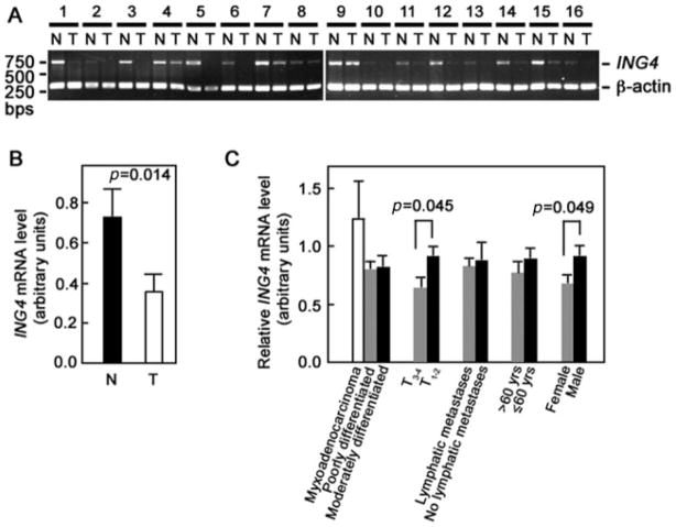
Figure 1.
ING4 mRNA is significantly reduced in gastric adenocarcinoma tissues compared with normal tissues. (A) Forty paired tumour and adjacent normal tissues were analysed for ING4 expression by RT-PCR. Two gels showing 16 representative paired tumour (T) and normal (N) tissues are shown. β-actin was used as internal loading control. (B) Average ING4 mRNA levels in 13 randomly picked paired tumour (T) and normal (N) samples were determined by real-time RT-PCR. (C) Analysis of relative ING4 mRNA level (T/N) according to gender (27 males, 13 females), age of patient (≤60 years, n = 23; >60 years, n = 17), lymphatic metastasis (lymphatic metastasis, n = 13; no lymphatic metastasis, n = 27), tumour stage (T1 – 2, n = 29; T3 – 4, n = 11), and extent of differentiation of the gastric adenocarcinoma (moderately differentiated, n = 10; poorly differentiated, n = 26; myxoadenocarcinoma, n = 4). Densitometry of PCR products was performed to analyse ING4 levels. Statistical analysis was performed by the t-test. p value <0.05 was considered significant

