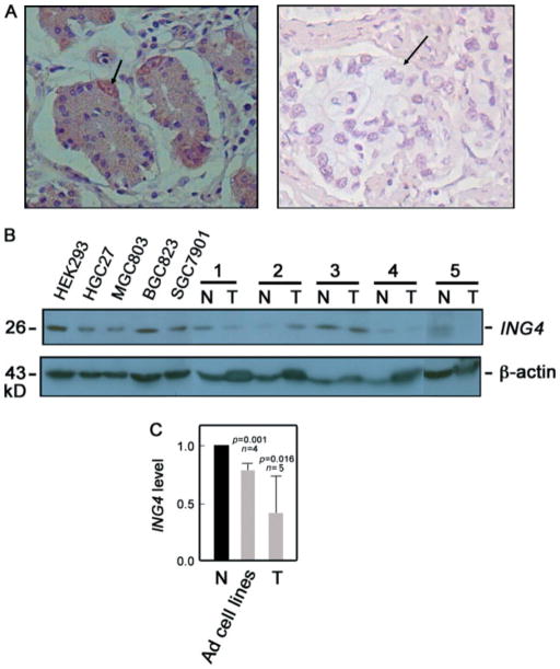
Figure 2.
ING4 protein expression is reduced in gastric adenocarcinoma cell lines and tissues. (A) ING4 expression in representative paired tumour (right panel) and normal adjacent (left panel) tissues was analysed by immunohistochemistry. Original magnification: 400 ×. Arrows indicate a stomach parietal cell (left panel) and a stomach adenocarcinoma nest (right panel). (B) Representative adenocarcinoma cell lines (HGC27, MGC803, BGC823, and SGC7901) and paired tumour (T) and normal (N) adjacent tissues (n = 5) were examined for ING4 protein expression by western blot analysis. HEK293 cells served as a positive control. β-actin blot was used for internal loading control. The 29 kD ING4 band detected in gastric adenocarcinoma tissues and cell lines is consistent with the possibility that the epitope recognized by the ING4 (T-15) antibody lies in an internal region of ING4 v1 and ING4 v2 but is absent or masked in the aberrantly spliced variant forms. (C) Analysis of the ING4/β-actin ratio following densitometric analysis of ING4 immunoblot. Values of normal tissues were normalized to 1. Statistical analysis was performed by the t-test tissue

