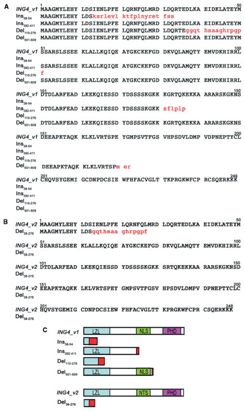
Figure 4.
Amino acid sequence alignment of the aberrantly spliced variant forms of ING4 v1 (A) and ING4 v2 (B) found in gastric adenocarcinoma tissues. Uppercase letters represent sequence identity, while lowercase letters in red indicate sequence variations due to a frame-shift in translation. (C) Diagrammatic representation of the aberrantly spliced variant forms of ING4 v1 and ING4 v2. Different conserved domains are indicated by different coloured boxes, including a leucine zipper-like motif (LZL); the nuclear localization sequence (NLS); and the plant homeo-domain (PHD). The red box indicates the region where sequence variations occur due to a frame-shift in translation

