
Fig. 3.
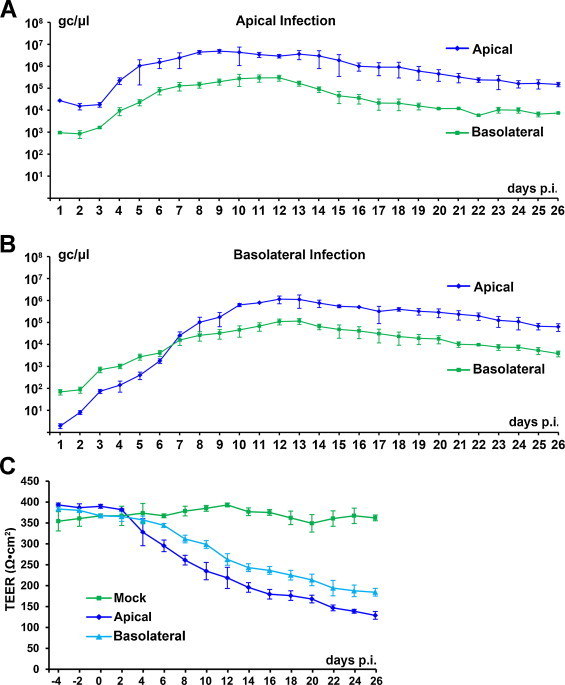
Kinetics of HBoV1-infected MucilAir HAE. MucilAir HAE was infected with HBoV1 from either the apical (A) or basolateral (B) surface at an MOI of 100 gc/μl. (A and B) At the indicated days p.i., progeny virions were collected from both the apical and basolateral chambers. HBoV1 virions were quantified by qPCR as viral gc/μl. Averages and standard deviations of the viral gc/μl are shown. (C) The TEER of HBoV1-infected HAE was monitored at the indicated days p.i. Averages and standard deviations of the detected TEER are shown.