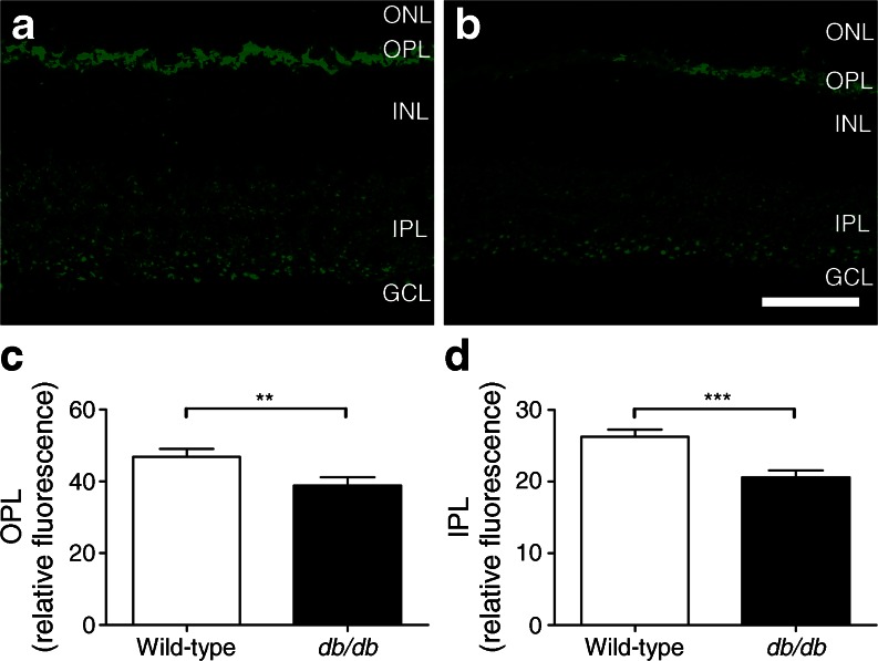
Fig. 8.
Altered retinal VGLUT1 immunoreactivity in diabetes. Retinas from (a) wild-type and (b) diabetic mice were assessed for VGLUT1 immunolabelling. The intensity of VGLUT1 labelling was assessed in (c) the OPL and (d) the IPL. In wild-type retinas, VGLUT1 labelling was strongly present in the OPL with punctate labelling in the IPL. The intensity of VGLUT1 labelling appeared decreased in diabetic retinas in both layers. Quantification of fluorescence intensity indicated significant decreases in both the OPL (**p < 0.01) and the IPL (***p < 0.001). Scale bar: 50 μm. GCL, ganglion cell layer; INL, inner nuclear layer; ONL, outer nuclear layer

