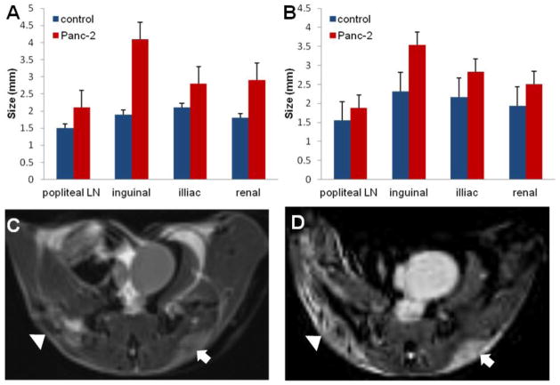
Figure 4.
Lymph node size measurements. The LN size measurements in vivo using HR-MRI (A) and ex vivo using a caliper (B) exhibited that the LNs in a normal mice were significantly smaller than those of the corresponding LNs in a PDAC tumor-bearing mice (all P-values were < 0.05, except in the popliteal LNs, where p>0.05). Representative T1W and T2W images of tumors in the same mouse are shown in C and D, respectively. MRI resolution is 0.14 × 0.14 × 0.14 mm3.

